baby chest x ray exposure
It is often the first type of imaging used to identify sources of pain evaluate traumatic injuries and locate a foreign body. Full legfull spine imaging is performed at 180 cm using CR.
Pathogens Free Full Text Chest Imaging For Pulmonary Tb Mdash An Update Html
Which is why getting a babys X-rays taken quickly and accurately is super important.

. X-rays use a small amount of radiation about the same levels that occur naturally in the environment. Routine prepost extubation X-rays 3. Your doctor can easily access these stored images to diagnose and manage your condition.
Full legfull spine imaging is performed at 180 cm using CR. If you recall from the chart a lower GI x-ray resulted in an exposure of 8mSv. X-ray examinations on the arms legs or chest do not expose your reproductive organs to the direct beam.
About one in eight scans ordered for kids is a CT scan. Irradiation of the testes and ovaries can cause infertility. High levels of acute exposure to radiation as low as 100 rad usually above 400 rad can cause acute radiation syndrome and even death.
The chin should not be superimposing any structures. It is during this period that the future vital systems of the future baby are formed. The greatest likelihood of developing complications occurs in the first trimester - that is the first 12 weeks of gestation.
Arms are not superimposed over lateral chest wall this can mimic pleural thickening. Exposure to radiation at a very young age is a pretty solid concern for both medical professionals and parents. Some conditions where X-rays are not indicated include.
Evaluation of an isolated episode of desaturation apnea 5. The only increased risk to these babies is a slightly higher chance of having cancer later in life less than 2 higher than the. Lung tissue absorbs little radiation and will appear dark on the image.
According to the Mayo Clinic radiation-induced health risks occur mostly at exposure levels of above 100mSv levels seen after nuclear meltdowns and atomic bombs. At Stanford we take extra precautions to minimize our patients exposure to radiation including using. At doses of around 10mSv so fairly close.
These higher levels have been shown to cause cancer. The most important downstream consequence specifically of X-rays for bronchiolitis is the inappropriate use of antibiotics Burstein says. It is intended to demonstrate an application of evidence-based research used in the creation of an exposure chart.
X-ray exams are used to help diagnose a wide variety of injuries and illnesses in children. With dental x-rays there is hardly any exposure to any part of the body except the teeth. On a chest x-ray the ribs and spine will absorb much of the radiation and appear white or light gray on the image.
Most x-ray images are electronically stored digital files. A certain danger is the X-ray of the ribs during pregnancy. Erect chest X-rays are taken at 180 cm.
Erect chest X-rays are taken at 180 cm. However x-rays of the torso such as the abdomen stomach pelvis lower back and kidneys have a greater chance of exposure to the uterus. The publication of this study and exposure chart could act as a benchmark for other medical imaging departments and to promote discussion on digital X-ray exposure optimisation for paediatrics.
A protective lead apron to shield certain parts of the body. Theres far less radiation exposure with an X-ray. Exposure to high-dose radiation two to eight weeks after conception might increase the risk of fetal growth restriction or birth defects.
Exposure to as little as 1 or 2 rad has also been associated with a slight increase in childhood malignancies especially leukemia. Lateral cervical spines are taken at 150 cm. Background Exposure groups based on.
Routine daily X-rays in ventilated neonates 2. X-rays are used throughout the body. All distal extremity exposures are taken at 110115 cm SID.
Patient position patient is supine. Ionizing radiation has several biological effects on reproduction. A chest X-ray is a painless noninvasive procedure with few risks.
Indications for a chest x-ray are far and wide from suspected infection to a pneumothorax. When an unborn baby is exposed to large doses of radiation above the dose received from 500 chest x-rays during the more sensitive stages of development. All distal extremity exposures are taken at 110115 cm SID.
During most x-ray examinations - like those of the arms legs head teeth or chest - your reproductive organs are not exposed to the direct x-ray beam. After re-intubation in a neonate where the optimal tip-to-lip distance is known based on initial X-ray 4. Because they spin around the body taking multiple images CT scans can deliver radiation doses that are up to 200 times higher than an.
The specific reason why these radiographs are conducted supine is the fact neonates will often stay on the ward performed mobile and do not have reliable head control until around 3 months old 13. 35 cm x 43 cm or 43 cm x 35 cm. However these dose levels arent used in diagnostic imaging.
Large radiation doses to the unborn baby during the more sensitive stages of development between weeks 2 and 15 of pregnancy can cause birth defects especially to the brain. The entire lung fields should be visible from the apices down to the lateral costophrenic angles. Chest x-ray is the most commonly used imaging exam for evaluating the chest.
In this study the exposure technique of 65 kVp and 16 mAs was chosen as a reference image due to this technique being near the suitable exposure uses. Lateral cervical spines are taken at 150 cm. Most researchers agree that babies who receive a small dose of radiation equal to 500 chest x -rays or less at any time during pregnancy do not have an increased risk for birth defects.
This can be either temporary or permanent depending on the dose as low as 15. Exposure to extremely high-dose radiation in the first two weeks after conception might result in a miscarriage.

X Ray For Kids Children S Health Orange County
Diagnostics Free Full Text A Pictorial Review Of The Role Of Imaging In The Detection Management Histopathological Correlations And Complications Of Covid 19 Pneumonia Html

Mengenal Cara Kerja Pemeriksaan X Ray Dan Efek Sampingnya Alodokter

Interpretation Of Neonatal Chest Radiography

Pediatric Chest X Ray In Covid 19 Infection European Journal Of Radiology

Neonatal Pneumonia Radiology Reference Article Radiopaedia Org

Indications For Chest X Rays In Children And How To Obtain And Interpret Them

What Is An X Ray For Kids Radiology And Medical Imaging

Reliability Of Chest Radiograph Interpretation For Pulmonary Tuberculosis In The Screening Of Childhood Tb Contacts And Migrant Children In The Uk Clinical Radiology

Pem Pearls Chest Radiographs For Shortness Of Breath
Interpretation Of Neonatal Chest Radiography

Interpretation Of Neonatal Chest Radiography

X Rays And Unshielded Infants Raise Alarms The New York Times

Interpretation Of Neonatal Chest Radiography
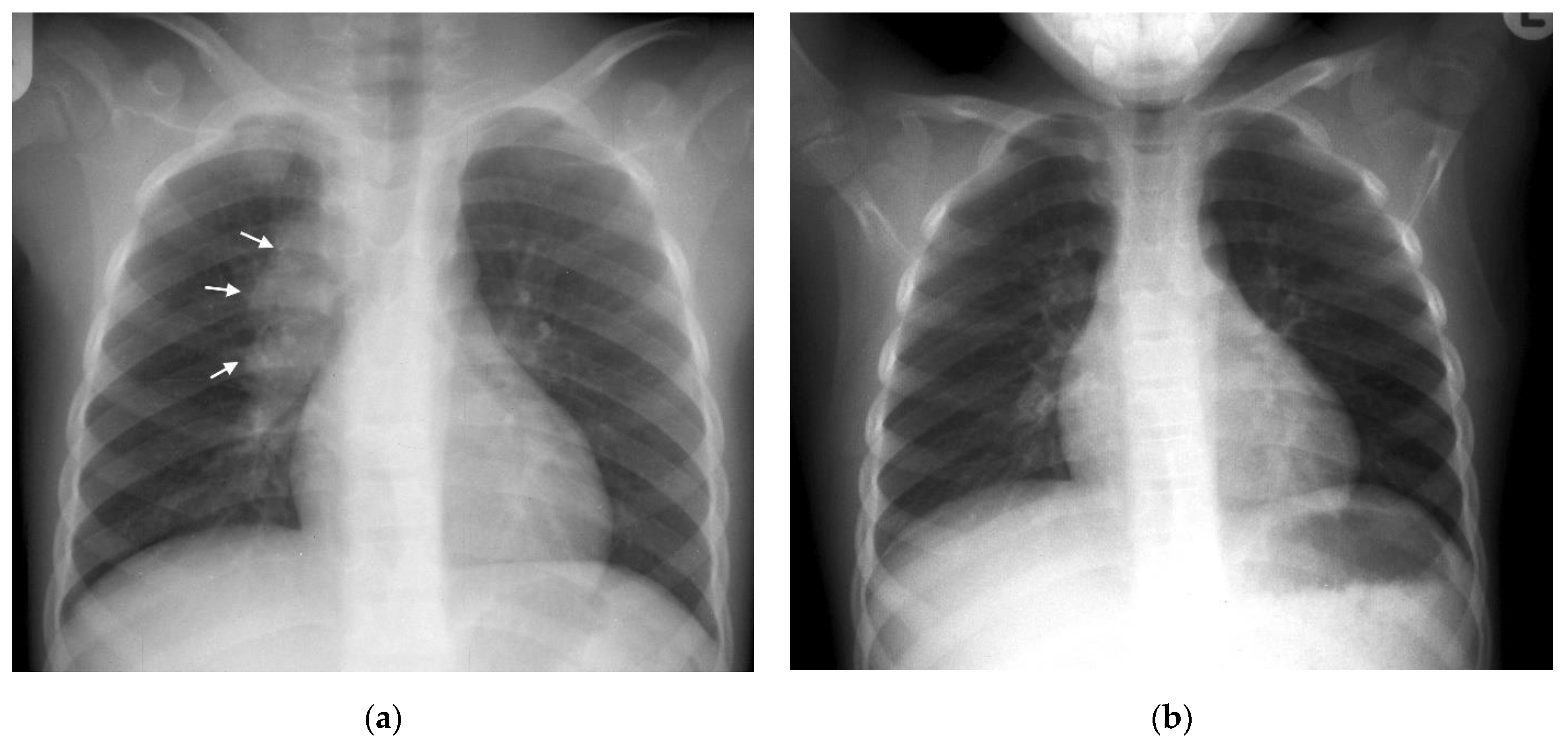
Pathogens Free Full Text Chest Imaging For Pulmonary Tb Mdash An Update Html

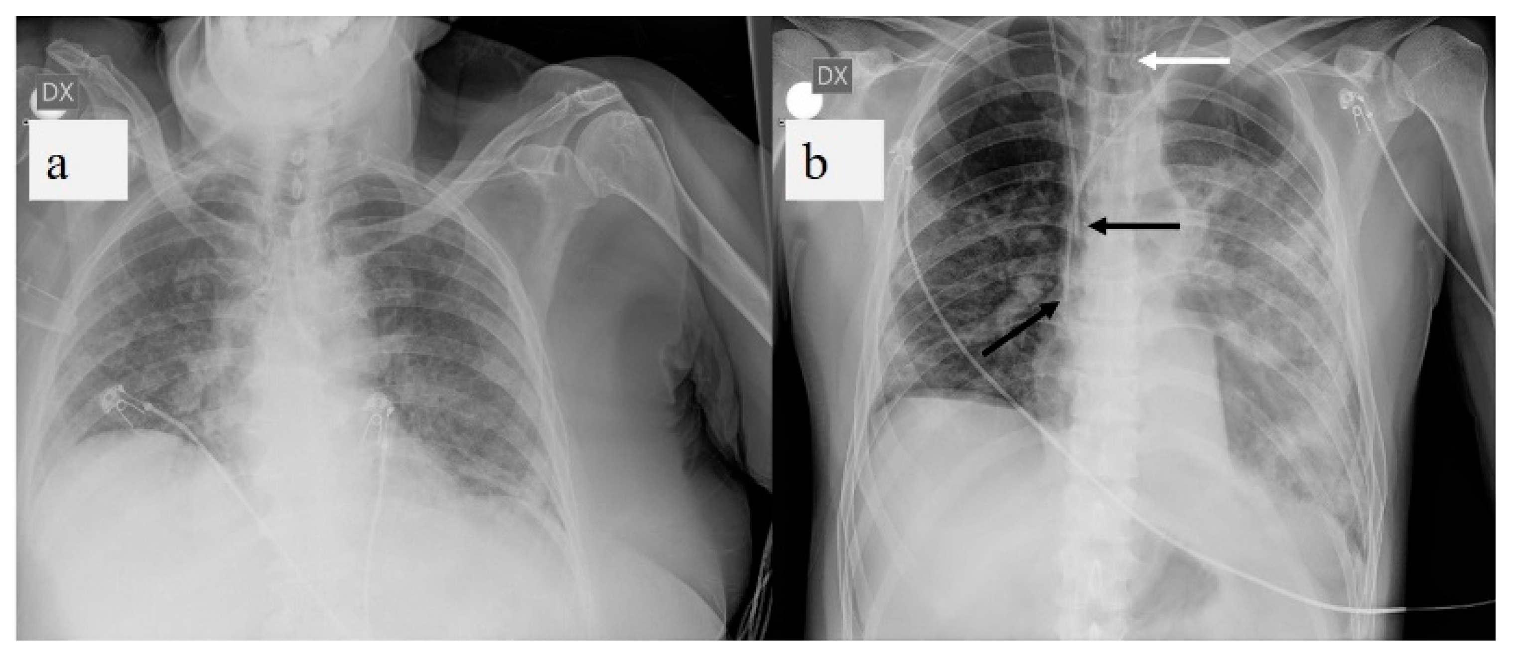
Pediatric Chest X Ray In Covid 19 Infection European Journal Of Radiology

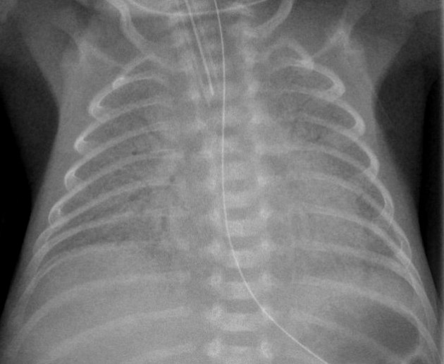
Neonate Chest Supine View Radiology Reference Article Radiopaedia Org
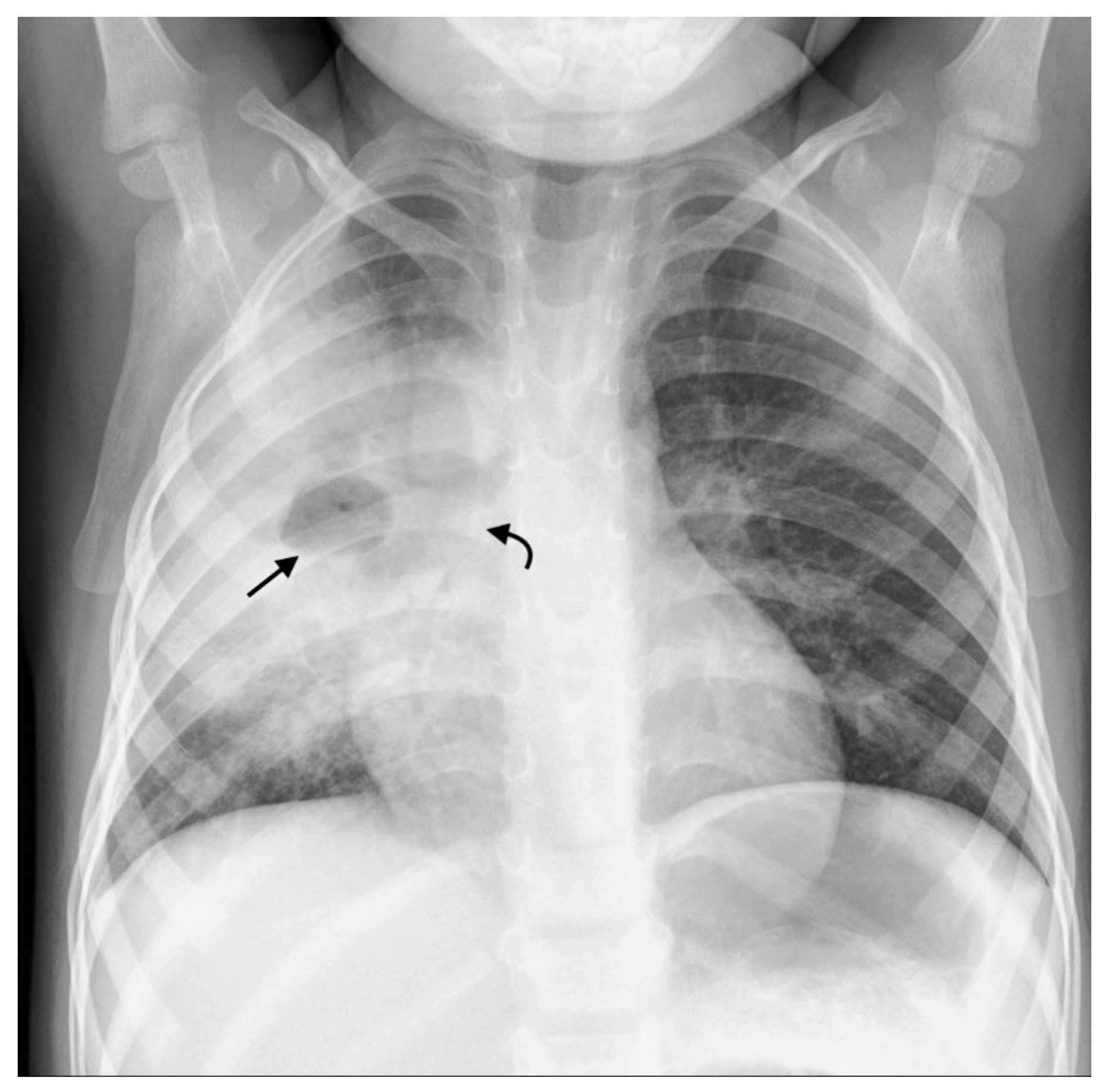
Neonatal Pneumonia Radiology Reference Article Radiopaedia Org

Neonatal Pneumonia Radiology Reference Article Radiopaedia Org